Urgent Care & Walk-ins are Welcome!

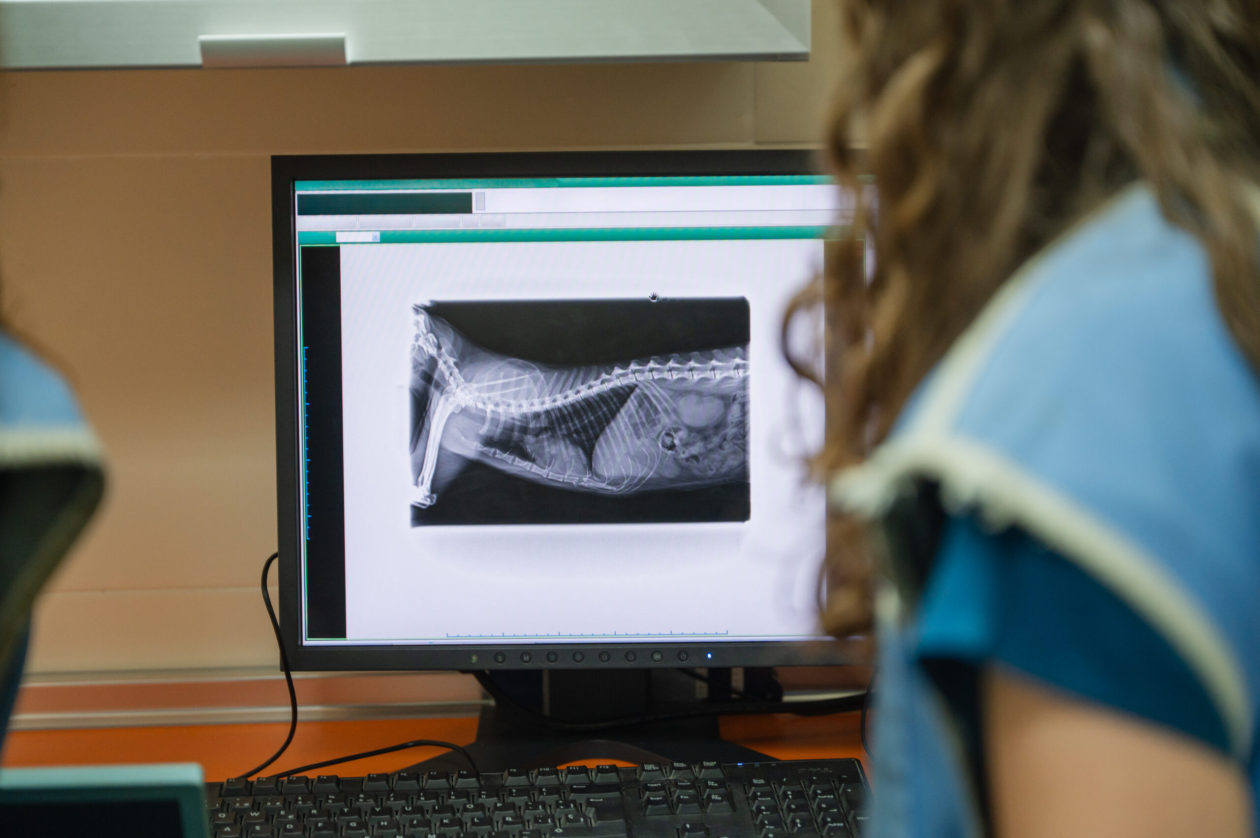
When a pet develops an unexplained health concern, a physical examination is often the starting point. Your veterinarian may listen to the heart and lungs, examine the mouth, feel the abdomen, evaluate movement, and ask about changes in appetite, behavior, or activity.
Sometimes, however, an examination cannot provide the complete picture. Certain problems involve structures inside the body that cannot be evaluated fully from the outside. This is where digital radiographs, commonly called pet X-rays, can become a valuable part of veterinary diagnostics.
Digital radiographs allow veterinarians to create images of internal structures and use those images alongside the physical examination and other diagnostic information. For pet owners in Bentonville, understanding when and why these images may be recommended can make the diagnostic process easier to understand.
What Are Digital Radiographs?
Digital radiographs use X-rays to create images of structures inside your pet’s body. The images can provide information about bones, the chest, the abdomen, and certain other structures.
Unlike a physical examination, which relies on observation and touch, radiographs give the veterinary team an internal view of areas that cannot be directly examined.
Digital technology also allows images to be produced and reviewed electronically. Your veterinarian can evaluate the images as part of the overall diagnostic process and determine whether additional evaluation may be appropriate.
Radiographs are not designed to replace a physical examination. Instead, they provide another source of information that can help answer specific questions about your pet’s health.
Why Would a Veterinarian Recommend an X-Ray?
There are many reasons a veterinarian may recommend digital radiographs. The decision generally depends on your pet’s symptoms, physical examination findings, medical history, age, and overall health.
For example, an X-ray may be considered when a pet has difficulty walking, persistent coughing, unexplained abdominal symptoms, or a suspected injury.
Radiographs can also be useful when the veterinarian needs to investigate an area more closely after noticing something unusual during the examination.
The goal is not simply to take an image. The goal is to obtain information that can help the veterinarian understand what may be happening inside your pet.
Evaluating Bones and Joints
Radiographs are particularly useful for evaluating bones and many joint structures. If your pet is limping, reluctant to move, or showing discomfort after an injury, your veterinarian may recommend X-rays.
The images can help identify fractures, changes in bone structure, joint abnormalities, and other concerns.
A pet may sometimes experience a movement problem without an obvious external injury. Radiographs can provide additional information that helps the veterinarian determine whether the problem involves the skeletal system.
This can be especially helpful when developing a treatment or monitoring plan.
Looking at the Chest
Digital radiographs can also provide useful information about the chest. Veterinarians may recommend chest X-rays when a pet has respiratory changes, persistent coughing, unusual breathing patterns, or other symptoms that warrant further evaluation.
Chest radiographs can allow the veterinary team to examine the lungs, heart silhouette, and other structures within the thoracic cavity.
They can help identify patterns that may not be apparent during a physical examination alone. However, radiographs are interpreted alongside other findings because an image by itself does not always provide a complete diagnosis.
Examining the Abdomen
The abdomen contains several important organs and structures that can be difficult to assess through physical examination alone.
When a pet experiences ongoing digestive symptoms, abdominal discomfort, changes in appetite, or other concerning signs, your veterinarian may consider abdominal radiographs as part of the diagnostic process.
Depending on the situation, X-rays may provide information about the size, position, or appearance of certain structures. They may also help identify patterns involving gas or other material within the digestive tract.
In some cases, another form of imaging may provide more detailed information. Your veterinarian can determine which diagnostic approach is most appropriate for the question being investigated.
X-Rays Can Support a Larger Diagnostic Plan
One of the most important things to understand about pet diagnostics care is that veterinarians rarely rely on one piece of information alone.
A physical examination provides information about your pet’s current condition. Blood or urine testing can provide insight into internal processes. Radiographs can show certain structures inside the body, while other imaging methods may provide different types of information.
These tools can complement one another.
For example, if laboratory findings raise a question about your pet’s health, your veterinarian may recommend imaging to gather additional information. Similarly, an abnormal finding on an X-ray may lead to additional testing.
The purpose is to connect the available information and develop a clearer understanding of the patient’s condition.
When an X-Ray Is Not the Only Option
Radiographs are valuable, but they are not appropriate for every diagnostic question. Different imaging methods provide different types of information.
For example, ultrasound can provide real-time images of many soft tissues and internal organs. Depending on the suspected condition, your veterinarian may recommend ultrasound, radiographs, laboratory testing, or a combination of diagnostic methods.
The choice depends on what the veterinary team needs to learn.
Rather than asking whether one test is better than another, it is more useful to ask what question the veterinarian is trying to answer. The appropriate test is the one that can provide relevant information for that particular situation.
Preparing Your Pet for Digital Radiographs
Preparation varies depending on the area being examined and the circumstances of the patient. Your veterinary team will explain any instructions that apply to your pet before the procedure.
Some pets may remain relatively calm during imaging, while others may be anxious or uncomfortable. Movement can affect image quality, so the veterinary team will work to position your pet appropriately.
In certain circumstances, sedation may be considered based on the pet’s temperament, comfort level, the type of imaging needed, and other medical factors. Your veterinarian can explain whether this may be appropriate for your pet.
What Happens After the Images Are Taken?
Once the radiographs are produced, the veterinary team evaluates the images together with your pet’s history and physical examination findings.
The veterinarian may explain what the images show and whether the findings provide an explanation for your pet’s symptoms. Sometimes the images may be reassuring, while in other situations they may indicate that additional testing is appropriate.
It is important to remember that an X-ray does not always provide an immediate final answer. Some conditions require multiple pieces of diagnostic information before a clear conclusion can be reached.
Why Your Pet’s History Still Matters
Even a high-quality radiograph needs clinical context. Your veterinarian needs to know what symptoms your pet is experiencing, when they began, whether they are changing, and what was discovered during the physical examination.
For example, an image of the chest is interpreted differently depending on whether the pet has coughing, breathing changes, reduced activity, or another concern.
This is why your observations at home are an important part of veterinary diagnostics. Tell your veterinarian about changes in eating, drinking, movement, bathroom habits, sleeping, and behavior.
Small details can help the veterinary team understand the bigger picture.
Choosing the Right Diagnostic Approach
The decision to recommend an X-ray is based on the individual patient. Your veterinarian considers the symptoms, physical findings, medical history, age, and suspected health concern before selecting an appropriate diagnostic approach.
At Dogwood Trails Animal Hospital, pet owners in Bentonville can discuss their concerns with a veterinary team and learn why specific diagnostic tests may be recommended.
Understanding the purpose of a test can help you feel more informed about your pet’s care. If your veterinarian recommends digital radiographs, ask what they are looking for, what the images may reveal, and whether additional testing could be needed.
Make Pet Diagnostics Care Part of Your Pet’s Health Plan
Digital radiographs can provide valuable information that a physical examination cannot always reveal. From bones and joints to the chest and abdomen, X-rays can help veterinarians investigate specific health concerns and make more informed decisions.
They are one part of a broader approach to pet diagnostics care, working alongside physical examinations, laboratory testing, and other diagnostic tools when appropriate.
If your pet has developed unexplained symptoms, changes in movement, digestive concerns, or other health changes, consider scheduling an online appointment with Dogwood Trails Animal Hospital in Bentonville. A veterinary consultation can help determine whether digital radiographs or another diagnostic approach may be appropriate.
Schedule a Veterinary Diagnostic Consultation
Concerned about a change in your pet’s health? Schedule an online appointment or booking with Dogwood Trails Animal Hospital in Bentonville to discuss your pet’s symptoms with a veterinary professional.
A thorough evaluation can help determine whether veterinary diagnostics, including digital radiographs, may provide useful information about your companion’s condition.
Frequently Asked Questions
Q1. What can digital radiographs show in pets?
Ans: Digital radiographs can provide images of bones, joints, the chest, abdomen, and other structures. They may help veterinarians investigate injuries, movement problems, respiratory concerns, digestive symptoms, and other health issues.
Q2. Does every pet with a health concern need an X-ray?
Ans: No. The need for radiographs depends on the pet’s symptoms, physical examination findings, medical history, and the specific question the veterinarian needs to investigate.
Q3. Are pet X-rays different from other diagnostic tests?
Ans: Yes. Each diagnostic tool provides different information. Radiographs are useful for certain internal structures, while laboratory testing and other imaging methods can provide different types of information. Veterinarians may use several tools together.
Q4. Will my pet need sedation for digital radiographs?
Ans: Not every pet needs sedation. The decision depends on factors such as your pet’s temperament, comfort, ability to remain still, and the type of imaging being performed. Your veterinary team can explain what is appropriate.
Q5. What should I tell my veterinarian before my pet has an X-ray?
Ans: Share details about your pet’s symptoms, when they started, changes in appetite or behavior, medications, previous health concerns, and any recent injuries. This information helps your veterinarian interpret the images in the proper clinical context.